Healthy Heart Function
|
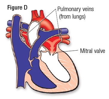
Normal blood flow through the heart begins with deoxygenated blood (depicted as blue) passing through the vena cava, from the body tissues, and into the right atrium of the heart. From there, atrial contraction pushes the blood through the tricuspid valve into the right ventricle (figure B). Blood then moves with low pressure through the pulmonary valve and to the pulmonary artery (figure C) which transports blood to the lungs where it becomes oxygenated. Oxygenated blood (depicted as red) now flows to the left atrium (figure D), and then, with atrial contraction, continues through the mitral valve to the left ventricle. Ventricular contraction pushes the blood through the aortic valve, into the aorta, and out to the body (figure E).
Ductus arteriosus- in a fetus, there is a passageway at the top of the descending aorta that connects it to the pulmonary artery. This allows blood to bypass the lungs, which are not yet operational, and continue on to circulate throughout the body. |

|


Retrieved from Retrieved from http://www.heart.org/HEARTORG/Conditions/CongenitalHeartDefects/AboutCongenitalHeartDefects/How-the-Healthy-Heart-Works_UCM_307016_Article.jsp#.WMPod4WcFPY
|
Transposition of the Great Arteries (TGA)
|
What is it?
Transposition of the great arteries (TGA) occurs when the pulmonary artery carrying blood to the lungs and the aorta, carrying blood to the body, switch positions (are transposed) in the heart. The pulmonary artery now rises from the left ventricle, while the aorta arises from the right ventricle. In some cases, these infants will also have a ventricular or atrial septal defect. It is usually detected prenatally or within the first few hours or weeks after birth, depending on the presence and size of septal defects. |
Retrieved from https://www.cincinnatichildrens.org/health/t/transposition
|
What causes it?
The causes for TGA are largely unknown, although some factors increase an infant's risk of developing this defect such as:
The causes for TGA are largely unknown, although some factors increase an infant's risk of developing this defect such as:
- history of rubella during pregnancy
- Poor nutrition during pregnancy
- alcohol consumption during pregnancy
- A mother older than age 40
- A mother who has poorly controlled diabetes
- Down syndrome in the baby
How does it effect the body?
Because these main arteries are transposed, the aorta carries unoxygenated blood from the right ventricle to the body, and back to the heart, without passing through the lungs. The pulmonary artery carries oxygenated blood from the left ventricle back to the lungs, and blood is then taken back to the left atrium without circulating the body. The body now depends on septal defects or a patent foramen ovale that allow blood to pass over to the left and right side of the heart to allow oxygenated and unoxygenated blood to mix which is then taken to circulate the body. If the defects are small, or not present, the infant is in severe danger of not receiving adequate oxygenation.
Because these main arteries are transposed, the aorta carries unoxygenated blood from the right ventricle to the body, and back to the heart, without passing through the lungs. The pulmonary artery carries oxygenated blood from the left ventricle back to the lungs, and blood is then taken back to the left atrium without circulating the body. The body now depends on septal defects or a patent foramen ovale that allow blood to pass over to the left and right side of the heart to allow oxygenated and unoxygenated blood to mix which is then taken to circulate the body. If the defects are small, or not present, the infant is in severe danger of not receiving adequate oxygenation.
 Retrieved from https://twohours.wordpress.com/category/transposition-of-the-great-arteries/ Truncus Arteriosus
How can I assess for it?
Infants with transposition of the great arteries will frequently present with:
cyanosis
oxygen saturation >90
rapid breathing
often, even in the presence of a septal defect, a murmur will be absent
lack of appetite
poor weight gain
Infants with transposition of the great arteries will frequently present with:
cyanosis
oxygen saturation >90
rapid breathing
often, even in the presence of a septal defect, a murmur will be absent
lack of appetite
poor weight gain
How is it treated?
Infants with transposition of the great arteries require surgery to reestablish normal blood flow to the body. Before surgery, doctors may give the infant prostaglandins to keep the ductus arteriosus open. This will allow blood to mix so that oxygen is able to get to the body. An atrial septostomy may also be performed prior to surgery in which a catheter is inserted to enlarge to foramen ovale connecting the atrium of the heart. This allows further mixing of blood resulting in additional oxygenation. Usually in the first few weeks of life, a surgery is performed to correct the defect called an arterial switch operation. In this surgery, the entry points for the aorta and pulmonary artery will be switched back to their proper place. Any septal defects that may also exist will be closed up at the time of the surgery.
Infants with transposition of the great arteries require surgery to reestablish normal blood flow to the body. Before surgery, doctors may give the infant prostaglandins to keep the ductus arteriosus open. This will allow blood to mix so that oxygen is able to get to the body. An atrial septostomy may also be performed prior to surgery in which a catheter is inserted to enlarge to foramen ovale connecting the atrium of the heart. This allows further mixing of blood resulting in additional oxygenation. Usually in the first few weeks of life, a surgery is performed to correct the defect called an arterial switch operation. In this surgery, the entry points for the aorta and pulmonary artery will be switched back to their proper place. Any septal defects that may also exist will be closed up at the time of the surgery.
