Healthy Heart Function
|
Normal blood flow through the heart begins with deoxygenated blood (depicted as blue) passing through the vena cava, from the body tissues, and into the right atrium of the heart. From there, atrial contraction pushes the blood through the tricuspid valve into the right ventricle (figure B). Blood then moves with low pressure through the pulmonary valve and to the pulmonary artery (figure C) which transports blood to the lungs where it becomes oxygenated. Oxygenated blood (depicted as red) now flows to the left atrium (figure D), and then, with atrial contraction, continues through the mitral valve to the left ventricle. Ventricular contraction pushes the blood through the aortic valve, into the aorta, and out to the body (figure E).
Ductus arteriosus- in a fetus, there is a passageway at the top of the descending aorta that connects it to the pulmonary artery. This allows blood to bypass the lungs, which are not yet operational, and continue on to circulate throughout the body. |


|


Retrieved from http://www.heart.org/HEARTORG/Conditions/CongenitalHeartDefects/AboutCongenitalHeartDefects/How-the-Healthy-Heart-Works_UCM_307016_Article.jsp#.WMPod4WcFPY
|
Tetralogy of Fallot
|
What is it?
Tetralogy of Fallot (TOF) is a congenital heart disease comprised of four characteristic defects:
|

Retrieved from http://smmhc.adam.com/graphics/images/en/18088.jpg
|
What causes it?
Most of the time, the causes for TOF are unknown, though poor maternal nutritional intake, maternal viral illness, and chromosomal abnormalities may increase fetal risk of developing this disorder.
Most of the time, the causes for TOF are unknown, though poor maternal nutritional intake, maternal viral illness, and chromosomal abnormalities may increase fetal risk of developing this disorder.
|
Retrieved from https://www.cincinnatichildrens.org/health/t/tof
|
How does it effect the body?
The VSD allows blood to flow back from the left ventricle and into the right ventricle. The right ventricle has to work extra hard and becomes hypertrophied because of the increase in blood volume, and also because pulmonary valve stenosis makes it hard to pump blood into the pulmonary artery. The septal defect also makes it possible for unoxygenated blood to flow into the left ventricle which is then able to travel through the improperly positioned aorta, and taken to the body tissues. |
 Retrieved from https://medlineplus.gov/ency/imagepages/18134.htm
How can I assess for it?
Signs of TOF will vary depending on the degree of malformation, especially concerning the pulmonary stenosis. However, symptoms may include:
Signs of TOF will vary depending on the degree of malformation, especially concerning the pulmonary stenosis. However, symptoms may include:
- cyanosis
- Shortness of breath and rapid breathing, especially during feeding or exercise
- Loss of consciousness (fainting)
- Clubbing of fingers and toes — an abnormal, rounded shape of the nail bed
- Poor weight gain
- Irritability
- A heart murmur may or may not be present depending on the severity of blockage through the pulmonary valve.
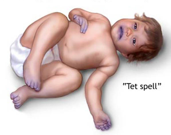
- tet spells- cyanosis accompanying crying or feedings, or periods of agitation in babies caused by rapid drops in oxygenation
- Pulse oximetry may be used to detect infants who experience mild hypoxia at rest, but do not display other manifestations during physical examination.
How is it treated?
Tetralogy of Fallot can only be corrected through surgery. There are two surgical options that may be considered. The most common option is to do a complete repair from the start, but sometimes a temporary shunt is placed to allow increased blood flow to the lungs in babies that are too weak for the complete surgery.
The complete intracardiac repair includes widening the pulmonary vessels and either widening or replacing the pulmonary valve. Surgeons will also place a patch over the VSD so that blood is no longer able to pass through and mix. Correcting these two defects solves the problems caused by an overriding aorta, and the issue of a hypertropic right ventricle.
