Healthy Heart Function
|
Normal blood flow through the heart begins with deoxygenated blood (depicted as blue) passing through the vena cava, from the body tissues, and into the right atrium of the heart. From there, atrial contraction pushes the blood through the tricuspid valve into the right ventricle (figure B). Blood then moves with low pressure through the pulmonary valve and to the pulmonary artery (figure C) which transports blood to the lungs where it becomes oxygenated. Oxygenated blood (depicted as red) now flows to the left atrium (figure D), and then, with atrial contraction, continues through the mitral valve to the left ventricle. Ventricular contraction pushes the blood through the aortic valve, into the aorta, and out to the body (figure E).
Ductus arteriosus- in a fetus, there is a passageway at the top of the descending aorta that connects it to the pulmonary artery. This allows blood to bypass the lungs, which are not yet operational, and continue on to circulate throughout the body. |


|


Retrieved from http://www.heart.org/HEARTORG/Conditions/CongenitalHeartDefects/AboutCongenitalHeartDefects/How-the-Healthy-Heart-Works_UCM_307016_Article.jsp#.WMPod4WcFPY
|
Hypoplastic Left Heart Syndrome
|
What is it?
Hypoplastic Left Heart Syndrome (HLHS) is a group of congenital malformations that involve the under-development of the left side of the heart (hypoplasia). These malformations may occur to differing degrees among different infants. They include a small, under-formed left atrium and ventricle, an abnormally narrow or closed mitral valve and aortic valve, and an abnormally narrow ascending aorta. Often, babies with HLHS will also have an Atrial Septal Defect, or a hole between the right and left atrium. What causes it? |
Retrieved from https://www.youtube.com/watch?v=3CP3xZVgpdg
|
It is not clear what causes HLHS. It is thought to be caused by a defect of a gene, chromosomal abnormality, or an environmental factor. Many times, it occurs along with other defects such as diaphragmatic hernia, omphalocele, and hypospadias.
How does it affect the body?
As the mitral valve is often severely malformed, blood is not always able to pass through it into the left ventricle. Instead, oxygenated blood passes through the Atrial Septal Defect, and back into the Right Atrium where it mixes with deoxygenated blood. It then flows to the Right Ventricle, which pumps blood back to the lungs, and the body through a Patent Ductus Arteriosus. Maintaining the Patent Ductus Arteriosus is therefore critical for the survival of the infant.
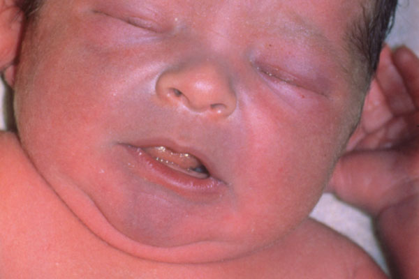
Retrieved from https://www.bing.com/images/search?view=detailV2&ccid=k%2FLOdgoN&id=D6E8935E37A06328AC09413B0E7D229D0D6C1158&q
=cyanotic+infant&simid=608034363927891256&selectedindex=4&mode=overlay&first=1&thid
=OIP.M93f2ce760a0d24fb8fd5222445027925o0
|
How can I assess for it?
Many times, HLHS can be diagnoses during an ultrasound while the infant is still in utero. After birth, HLHS causes symptoms similar to those found in other heart conditions. They may include:
|
How is it treated?
An infant will not be able to live for very long with this condition without medical intervention. Prostaglandins will be given to the infant after birth in order to keep the Ductus Arteriosus patent. The infant will need to undergo surgery to help increase blood flow, and bypass the left side of the heart. The surgery is performed in three stages:
An infant will not be able to live for very long with this condition without medical intervention. Prostaglandins will be given to the infant after birth in order to keep the Ductus Arteriosus patent. The infant will need to undergo surgery to help increase blood flow, and bypass the left side of the heart. The surgery is performed in three stages:
- Norwood Procedure- this procedure is typically performed in the first few weeks of an infant's life. It involves the creation of a new aorta that is connected to the right ventricle to allow for increased blood flow.
- Glen Shunt Procedure- this procedure is typically performed at 4 to 6 months of age. This procedure involves forming a connection between the superior vena cava and the pulmonary arteries. This allows deoxygenated blood to flow directly to the lungs, and takes some of the strain off of the right ventricle.
- Fontan Procedure-This procedure is typically performed when the child is between 18 months and 3 years old. This procedure connects the inferior vena cava to the pulmonary artery. This allows all deoxygenated blood to flow directly to the lungs, bypassing the heart. The Right ventricle is now only responsible for pumping blood to the body, and oxygenated and deoxygenated blood will no longer mix within the heart.
